


Technologies
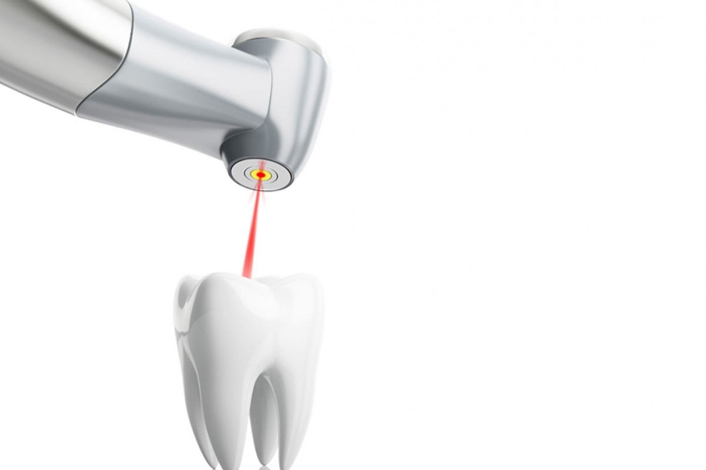
Orcos Medical - Laser Technology
Laser technology today represents an innovative and highly reliable tool, capable of ensuring superior quality treatments. It is effective in treating peri-implantitis, tooth sensitivity, cold sores and endodontic therapies.
Swiss Dental Clinic collaborates with Orcos, a young Swiss company and leader in the national market, specialising in laser technologies for dentistry and dermatology, both aesthetic and therapeutic.
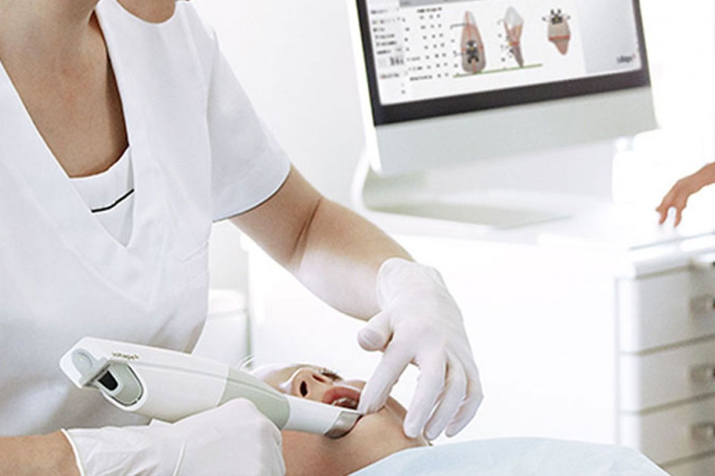
Digital Impression Planning
The intraoral scanner is a 3D device that precisely captures the shape and dimensions of the dental arches, transforming them into digital data to be displayed in real time.
Thanks to a light beam and high-resolution cameras, it allows the dentist to plan restorations, reconstructions or corrective appliances with maximum accuracy. Compared to traditional impressions, it is less invasive, eliminates sticky materials, reduces stress and discomfort, ensures precision even in the most hidden areas and speeds up data transmission to the dental laboratory.
The intraoral scanner is suitable for prosthetics on natural teeth and implants, orthodontic appliances for adults and children, pre- and post-treatment documentation, and support for guided surgery, offering a safe, efficient and innovative approach to every dental treatment.
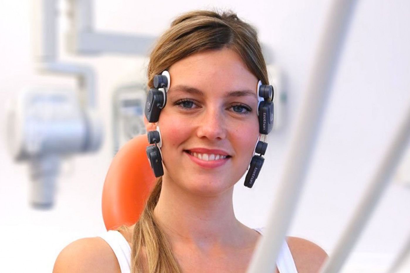
Teethan
In our Lugano practice, we use Teethan®, the first completely wireless medical device for the simple, rapid and non-invasive analysis of dental occlusion, the contact between the upper and lower arches during chewing.
Improper occlusion can affect chewing, swallowing, speaking and posture, causing muscle tension, dental problems, headaches and neck pain. Thanks to Teethan®, we can obtain precise information and numerical data on the patient's muscle activity and occlusal balance, allowing us to plan targeted corrective therapies.
The test lasts about 3 minutes, is painless and uses four small, lightweight probes applied to the temporalis and masseter muscles, which detect electrical signals during brief clenching trials. The result is a detailed analysis of muscle prevalence, torsion and imbalance, obtained quickly, safely and without any discomfort, ensuring patient comfort and the specialist reliable scientific data for effective intervention.
Intraoral X-Ray
An intraoral x-ray is a radiological examination that allows for an in-depth and immediate analysis of a specific area of the mouth, providing the dentist with all the information necessary for immediate intervention.
Using small x-rays, it is possible to evaluate the roots, gums, periodontal ligament and alveolar bone of one or two teeth, with precise and non-invasive results.
There are several types of intraoral x-rays: the bite-wing, useful for identifying cavities; the periapical, which shows the entire length of the tooth from crown to root and is indicated for granulomas or abscesses; and the occlusal, larger, which allows for the visualisation of approximately three-quarters of the dental arch, offering a detailed and immediate analysis for any intervention.
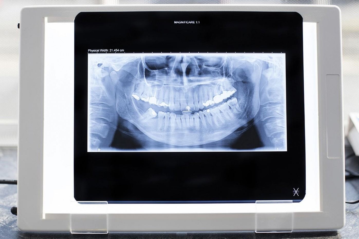
Panoramic X-Ray
Orthopantomography, or dental panoramic imaging, is a first-level examination that allows us to obtain a complete picture of the health of the stomatognathic system, allowing us to define the most appropriate treatment plan.
This exam produces detailed images of the teeth, dental arches and maxillary and mandibular bones, allowing the detection of cavities that are not visible to the naked eye, cysts, granulomas, root fractures and other abnormalities.
The dental panoramic scan also allows us to evaluate the alignment of the arches and the presence of structural problems, all in about a minute, without any preliminary preparation, making the examination quick, simple and precise.
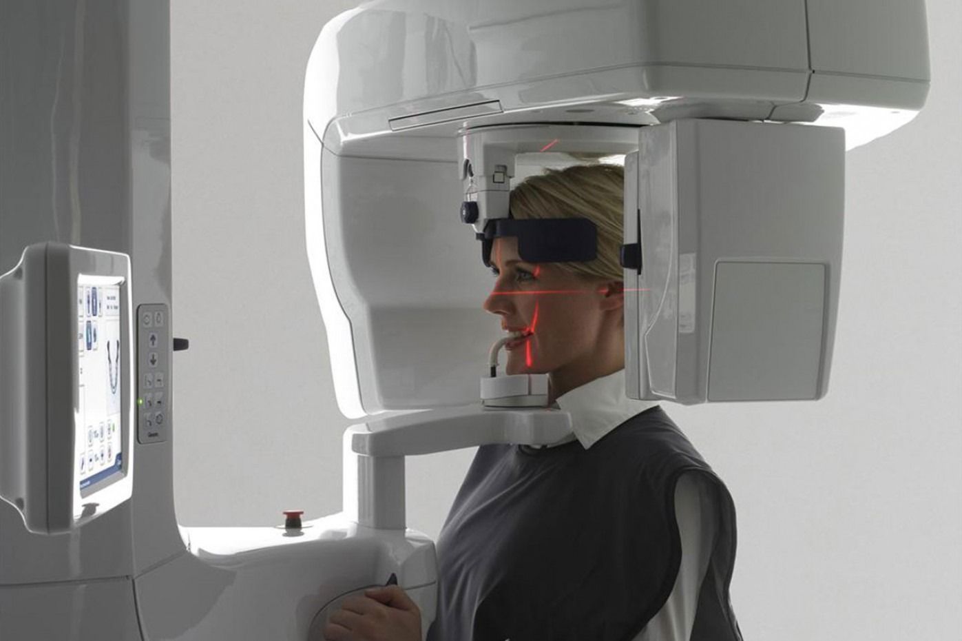
3D DentalScan
In our dental practice, we use high-precision diagnostic equipment, such as Digital Panoramic Imaging and DentalScan, to assess the health of the dental arch prior to surgery or implant placement. These technologies allow us to obtain detailed x-rays and CT scans, minimising radiation and ensuring a precise, three-dimensional view of the bone and teeth.
Panoramic dental x-rays provide a comprehensive overview of both jaws and the maxillofacial region, detecting cavities, cysts, or other issues that could affect the feasibility of the procedure. A 3D DentalScan CT scan allows for the assessment of bone quantity and quality, which is essential for complex implants and high-precision rehabilitation.
Thanks to digitalisation and the highest definition images, we can plan every treatment with absolute precision, identifying anomalies or pathologies with minimal margins of error, guaranteeing safety and effectiveness for our patients.